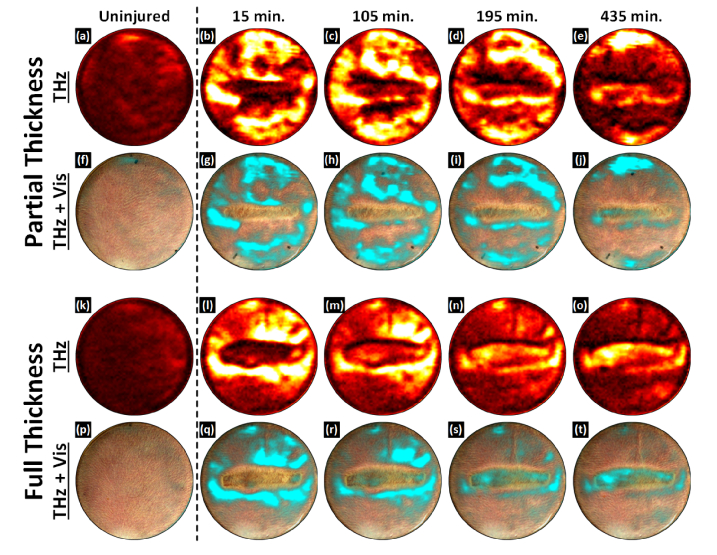
Fig. 7.
Burn wound time series imaging results. (a) – (e) THz images of a partial thickness burn. (f) – (j) Partial thickness THz images superimposed on the registered visible frame. (k) – (o) THz images of a full thickness burn. (p) – (t) Full thickness THz images superimposed on the registered visible frame. THz contrast is distinct for each burn wound severity. In time-series THz images of a partial thickness burn, the contact area shows a drop in TWC and an edematous front superior to the burn over time. In contrast to the partial thickness wound the contact area of a full thickness burn does not display a significant drop in TWC. Additionally, the contact area is surrounded by a ring of TWC which runs concentric and coincident with the burn contact zone.

