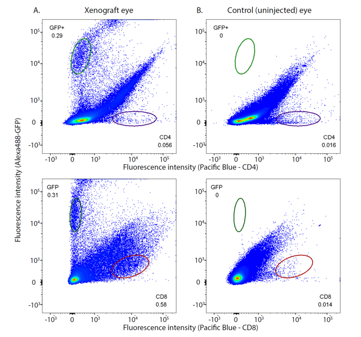
Fig. 6.
Flow cytometry of cells from an eye with a xenograft glioblastoma reveals a tumor-associated increase in cells expressing CD8 epitope. In all four panels the ordinate gives the fluorescence intensity of cells labeled with an anti-GFP antibody tagged with Alexa 488 dye, while the abscissa is the fluorescence intensity of cells labeled with antibodies to CD4 (upper row of panels) or CD8 (lower row) conjugated to Pacific Blue dye. The left column of panels was obtained with cells from the eye that had a glioblastoma xenograft, while the right column of panels was obtained with exactly the same gating parameters from the cells identically harvested from the control (uninjected) eye of the same nude mouse. Counts in the plot regions indicated by the ellipses were used for statistical analysis. There were no GFP+ counts in the cells from the uninjected eye. The eye with the xenograft had statistically reliable 3.5-fold and 41-fold increases in cells expressing CD4 and CD8 cell surface markers, respectively, relative to the control eye. The flow cytometry data were analyzed with FloJoTM software and the numbers on the panels represent the fractions of the total count in the elliptical regions. The Type 1 tumor was generated by injection of 500 cells; the corresponding growth curve is illustrated in Fig. 3A by filled orange circles. This experiment was replicated in a second mouse with similar results.

