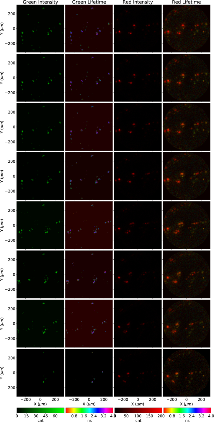
Fig. 7.

Sequence of eight images extracted from a ten-frames, 1 FPS FLIM movie. Microspheres covalently bound to Fluorescein (green), TAMRA (orange), NBD (yellow) and CY5 (red) dyes were deposited in a microscope slide. The imaging fiber was mounted on a XYZ translation stage and moved a few across the surface of the slide.
