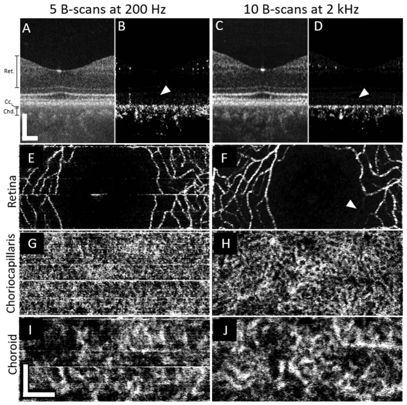
Fig. 3.
A direct comparison of the imaging performance of the two swept-source systems of a foveal scan from subject 4, a 36-year-old emmetropic male. Images from the slow system are shown on the left half (A,B,E,G,I) of the figure while the right half (C,D,F,H,J) shows images produced using the fast system. En face images from the fast system (right side) have been cropped, height-wise, to match the corresponding images from the slower system (left side). White arrows in B and D show that the fast system suppresses bulk motion-related noise in the vessel-free region of the pigment epithelium and photoreceptors better than the slow system. A white arrowhead in F shows that a this specific retinal capillary appears fainter in the fast system, most likely due to a momentary reduction of flow during the short period of scanning. All scale bars are 200µm.

