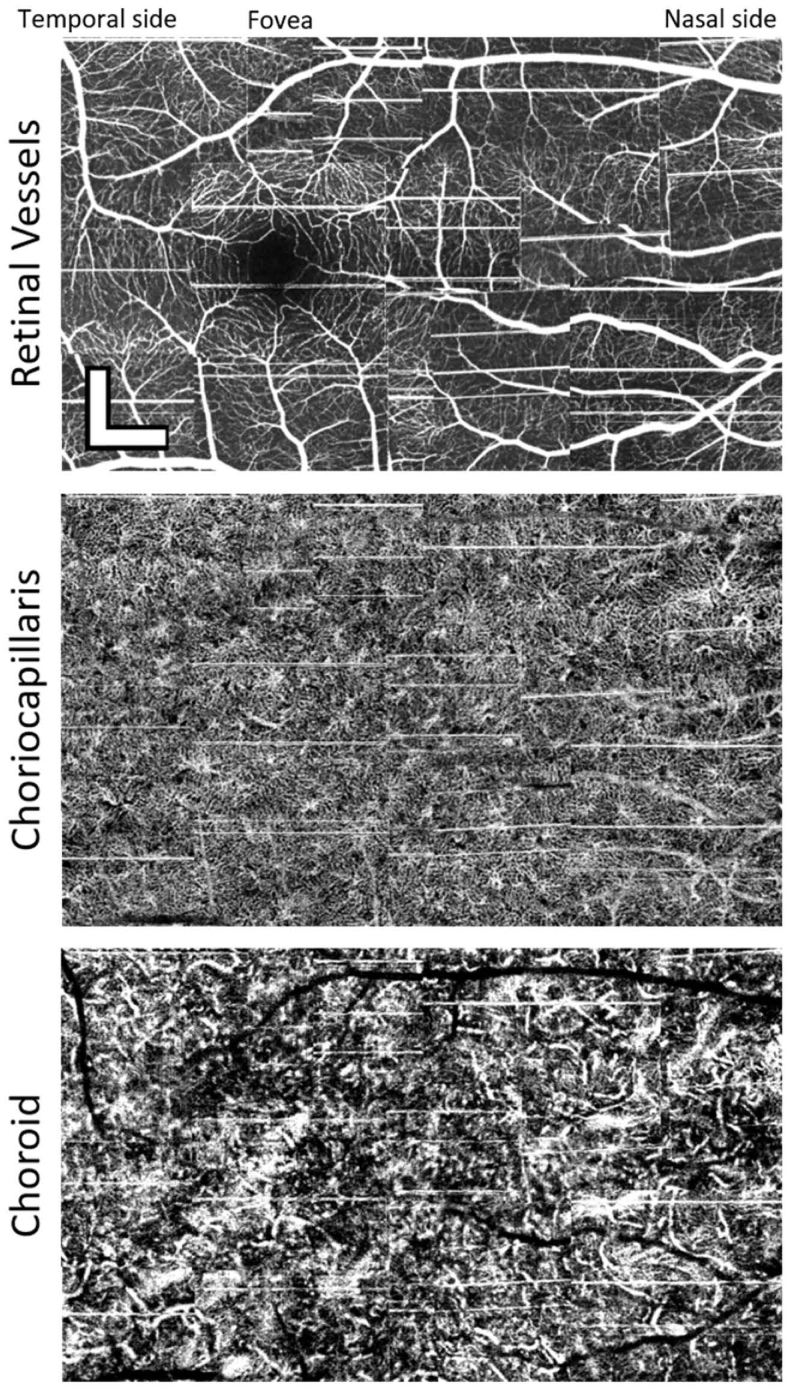
Fig. 6.
Example of a mosaic of 15 images taken from the right eye of Subject 1, a 32-year-old male with 2.5 diopters of myopia. The images were taken with the high-speed system. The first mosaic shows the maximum intensity projection of all the outer retinal layers. The second panel shows a mean of the projections within the range of the choriocapillaris. The bottom mosaic shows a mean projection of the choroid. Depth ranges of each projection are the same as those indicated by the annotations to the left of Fig. 4(A). Images are stitched together manually without any intra-volume motion correction. Scalebars are 300µm.

