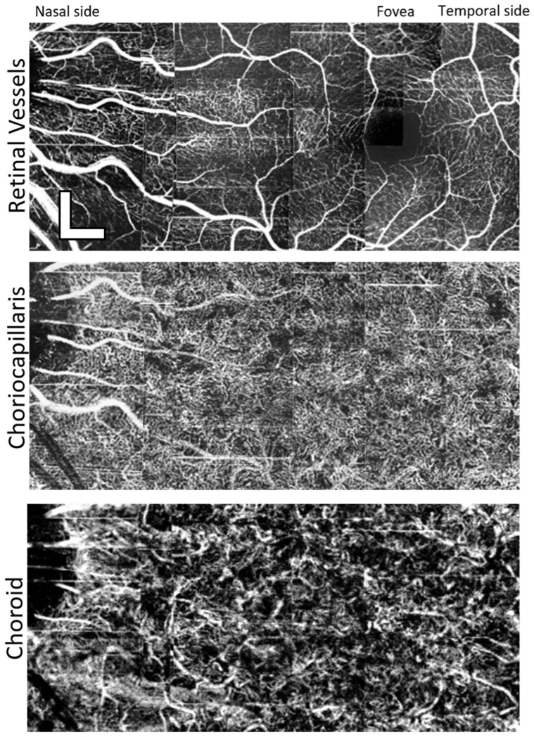
Fig. 7.
Example of a mosaic of 18 images taken from the left eye of Subject 4, a 36-year-old emmetropic male. The images were taken with the high-speed, 1.64 MHz system. The images were assembled in the same manner as for Fig. 6. It is noteworthy that the choriocapillaris appears so have different vessel densities between the two subjects. Scalebars are 300µm.

