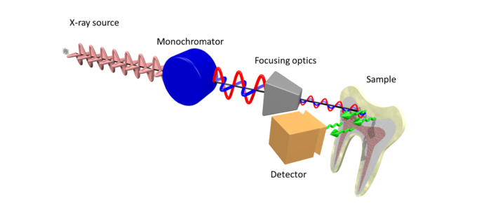
Fig. 1.
Schematic illustration of the main optical components of the setup installed at beamline ID21 for XRF-PIC mapping. Two undulators are used to generate the X-rays that are polarized in the horizontal plane, subsequently monochromatized by a fixed exit double-crystal Si(111) Kohzu-monochromator, then focused using KB optics. The polarization orientation is not affected by any of the components downstream of the source until impinging on the sample. Following interaction with the apatite (in the tooth), non-polarized XRF radiation is generated. The total XRF signal (illustrated by green arrows) is collected by a photodiode detector.

