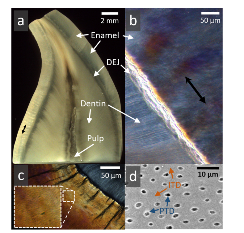
Fig. 2.
Typical human tooth tissues (a). Cross sectional slice in a human canine crown revealing the outer enamel (worn on the upper left edge) encasing a large bulk of dentine. These 2 tissues are attached at the dentine-enamel junction (DEJ). The pulp is partially stained. (b) Higher magnification of a segment of enamel, DEJ and dentine, viewed in polarized light. Groups of alternating prism orientations in enamel (black double headed arrow) exhibit Hunter Schreger optical bands. (c) In cross section, enamel surrounds dentine (upper right) attached at the DEJ where dark tufts of organic material extend outwards (magnified area shows polarization light microscope image of clusters of enamel prisms). (d) Typical backscattered electron microscopy image of dentine containing tubules, perforating the intertubular (ITD) tissue. Tubules lie approximately 10 µm apart and are ~1 µm in diameter. Many tubules are lined by a thin highly-mineralized sheath of peritubular dentine (PTD) within ITD.

