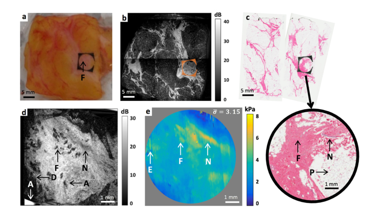
Fig. 8.
Excised human breast tissue. (a) Specimen photograph with the fiducial marker in place. (b) Wide-field OCT image taken with a benchtop scanning head. The position of the fiducial marker has been overlaid (in orange). (c) Histology with the inset highlighting the region scanned by OCT. (d) Motion-corrected en face OCT image at ∼0.1 mm below the tissue surface (e) Motion-corrected optical palpogram. The area outside of the fiducial marker has been masked in grey. is the average stress in kPa, and equivalently, pressure applied to the tissue during the scan. A, interpolation inaccuracy artifact. D, field curvature artifact. E, stress estimation artifacts. F, fibrosis from previous surgery. N, nerve. P, adipose.

