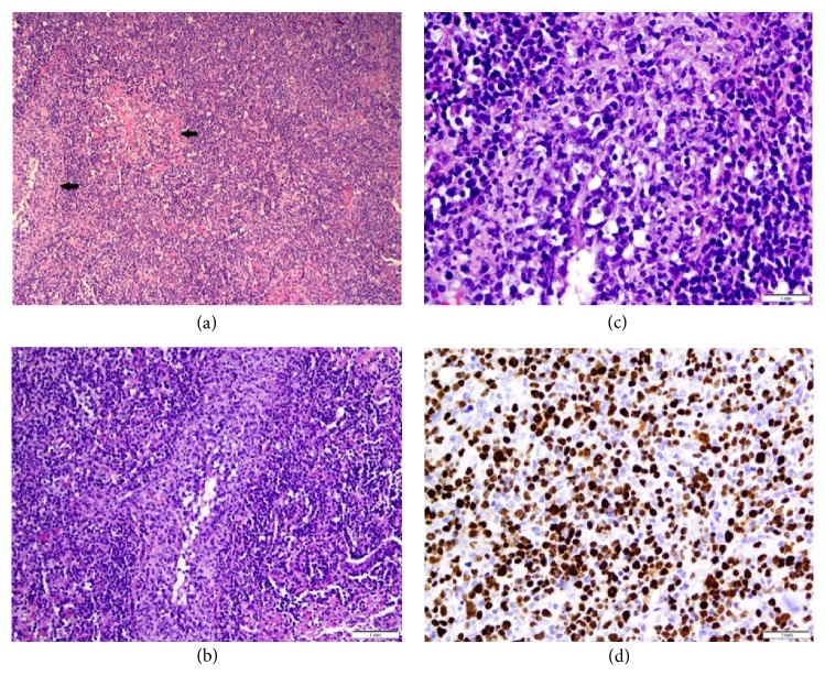
Figure 2.
Low power section (a) from lung biopsy showing a diffuse neoplastic lymphoid cells (arrows) infiltrating the lymphatic and bronchovascular bundles. (H&E, 100x) Medium power view (b) showing neoplastic cells involving walls of pulmonary vessels. Small, reactive lymphocytes are seen in the background. (H&E, 200x) High power view (c) shows large, atypical B-cells with irregular nuclear membranes, open chromatin, and prominent nucleoli invading vessel walls (angioinvasion). (H&E, 500x) The neoplastic B-cells were positive for EBV (d).

