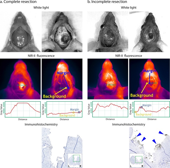
Figure 3.
For both Complete resection (a) and Incomplete resection (b), left-hand side pictures are taken prior to tumor resection and right-hand side pictures are after resection. Complete resection: no signal left around the tumor at the postoperative NIR-II picture and it is considered a complete resection. Yellow circle indicates the margin (M = 36,000 Gray Value) and the blue circles indicates the tumor free background signal (B = 35,000 Gray Value) resulting in a margin-to background ratio = 1.0. Incomplete resection: clear signal at the tumor margin at the postoperative picture indicating positive resection margins. Yellow circle indicates the positive margin (M = 52,500 Gray Value) and the blue circles indicates the tumor free background signal (B = 35,000 Gray Value) resulting in a margin-to-background ratio = 1.5. On ICH, blue arrows are pointing at uPAR positive stained tumor cells.

