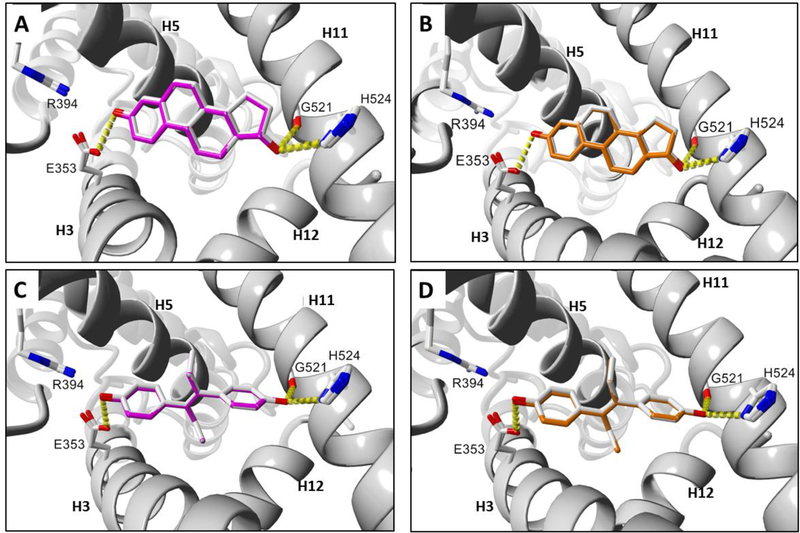
Fig. 4.
Docking comparisons of known ERα agonist ligands in human and rodent ERα-LBD receptors. (A) E2 human and mouse. (B) E2 human and rat. (C) DES human and mouse. (D) DES human and rat. Ligand colors: gray = human, magenta = mouse, orange = rat. Hydrogen bonds are represented as yellow dashes. Oxygen and nitrogen atoms are colored red and blue, respectively. Active site helix labels (H5, H11, and H12) are displayed in bold face. Hydrogen-bonding residues are displayed and labeled.

