Figure 3.
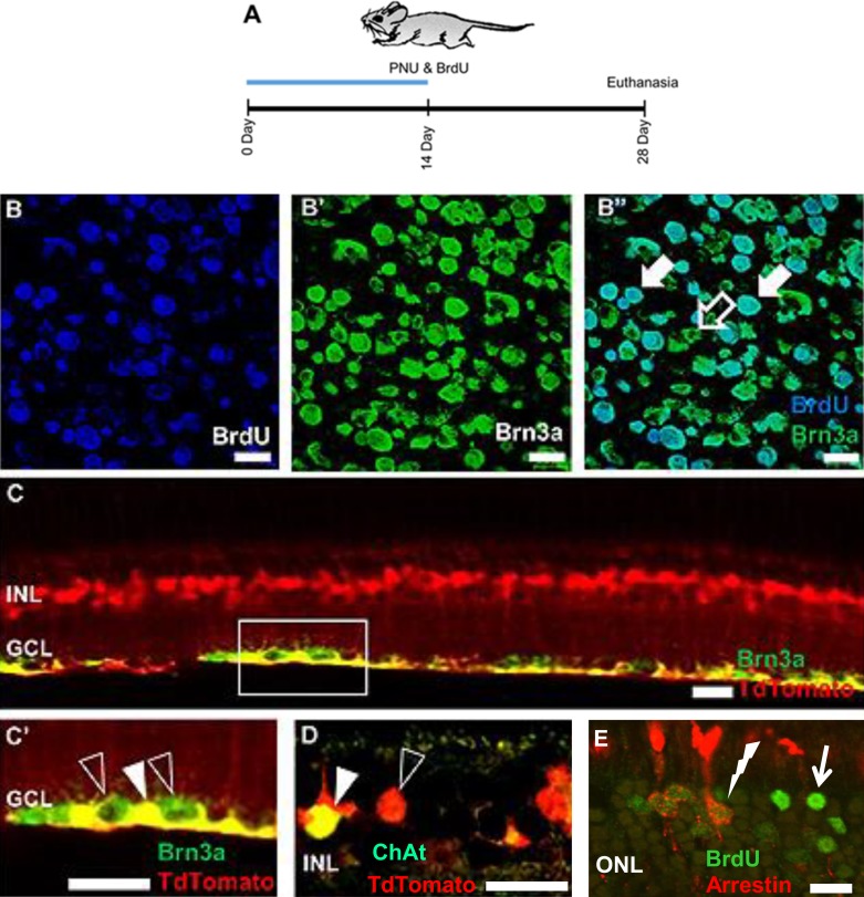
Genesis of tdTomato-positive adult retinal neurons following PNU-282987 treatment. Confocal images from adult transgenic mice that were treated with PNU-282987 for 2 weeks and euthanized 4 weeks after the start of treatment (A). RGCs (B–B” and C–C') and starburst amacrine cells (D) showed colabeling for the Müller glia marker tdTomato. In addition, cone photoreceptors double-labeled with antibodies against BrdU and arrestin (E). The open arrow in (B” ) indicates a Brn3a+, BrdU− RGC, whereas closed arrows show cells in the GCL double-labeled with antibodies against both Brn3a+ and BrdU. The open arrowhead in (C') shows Brn3a+ RGCs, whereas the closed arrowhead illustrates a double-labeled Brn3a+, tdTomato+ RGC. The open arrowhead in (D) illustrates cell bodies of Müller glia in the INL (red), whereas the closed arrowhead indicates a ChAT+, tdTomato+ starburst amacrine cell. In (E), the solid arrow points to a BrdU+ cell in the ONL, whereas the lightning bolt illustrates a double-labeled cell in the ONL labeled with antibodies against arrestin and BrdU. Scale bars: 20 μm. n = 6 mice for each experimental condition.

