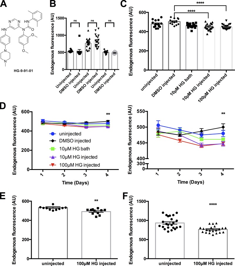
Figure 2.
Addition of the SIK inhibitor decreases background fluorescence. (A) Chemical structure of the small-molecule SIK inhibitor HG 9-91-01. (B) Comparison of endogenous fluorescence between several batches of uninjected and 0.5% DMSO-injected oocytes. A one-way ANOVA with a post hoc Holm–Sidak multiple comparisons test found no significant difference compared with uninjected oocytes (uninjected: n = 11, 19, and 11; DMSO: n = 10, 18, and 11). For further information, see Table S3. (C) Effect of HG 9-91-01 endogenous fluorescence under different conditions: uninjected (circles, n = 14), 0.1% DMSO injected (diamonds, n = 9), 10 µM HG 9-91-01 bath (squares, n = 22), 10 µM HG 9-91-01 injected (triangles, n = 21), and 100 µM HG 9-91-01 injected (upside-down triangles, n = 21) measured on d 4. ****, P < 0.0001 compared to uninjected oocytes, one-way ANOVA with a post hoc Holm–Sidak multiple comparisons test. For further information, see Table S4. (D) Endogenous fluorescence under different conditions measured over time, as described in B. Panel on the right is a zoom in to demonstrate the effect of the different conditions described in B. **, P < 0.001, 10 µM HG injected and 100 µM HG injected on day 4 (two-way ANOVA with a post hoc Dunnett multiple comparisons test). For further information, see Table S5. (E) Results from a darker batch of oocytes where HG 9-91-01 was tested (uninjected: n = 11; 100 µM HG 9-91-01 injected: n = 11). **, P = 0.0031, unpaired two-tailed t test. (F) Results from a lighter batch of oocytes where HG 9-91-01 was tested (uninjected: n = 22; 100 µM HG 9-91-01 injected: n = 14). ****, P < 0.0001, unpaired two-tailed t test. All error bars are ±SEM centered on the mean..

