Figure 5.
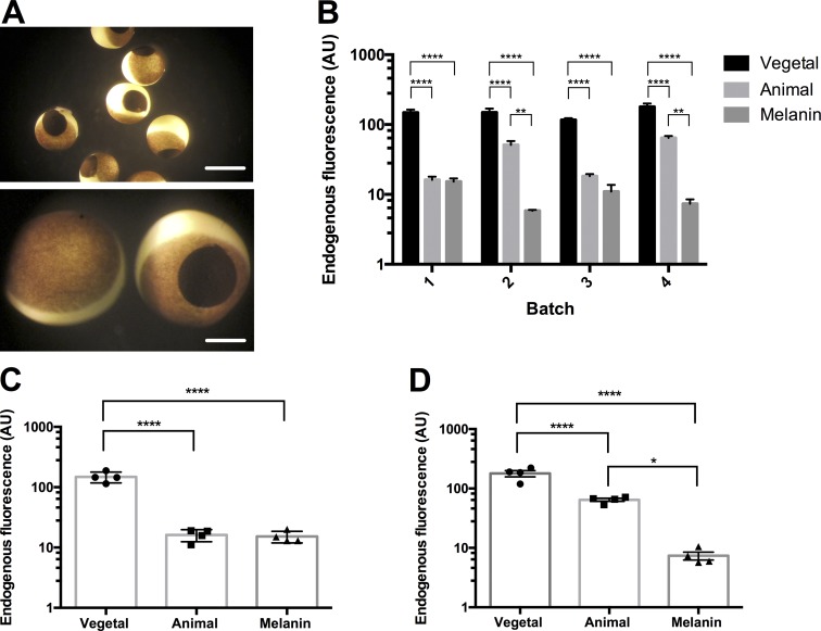
Synthetic melanin injections significantly improve fluorescence recording conditions. (A) Representative X. laevis oocytes injected with 50.1 nl of 30 mg/ml melanin. Scale bars, 1 mm (top) and 0.3 mm (bottom). (B) Improvement of endogenous background fluorescence of four separate batches of oocytes at 535 nm. Batch 1, n = 4; batch 2, n = 6; batch , n = 10; and batch 4, n = 4. *, P < 0.05; **, P < 0.01; ****, P < 0.0001, two-way ANOVA with a post hoc Tukey multiple comparison test. (C) The improvement in endogenous background fluorescence in “dark” oocytes, <20 AU at 535 nm (vegetal, n = 4; animal, n = 4; and melanin injected, n = 4). ****, P < 0.0001, ordinary one-way ANOVA with a post hoc Tukey multiple comparison test; no statistically significant difference was found between the animal pole and melanin. (D) The improvement in endogenous background fluorescence in “light” oocytes, >20 AU at 535 nm (vegetal, n = 4; animal, n = 4; and melanin injected, n = 4). *, P < 0.05; ****, P < 0.0001, ordinary one-way ANOVA with a post hoc Tukey multiple comparison test. For further statistical information, see Tables S13–S15.

