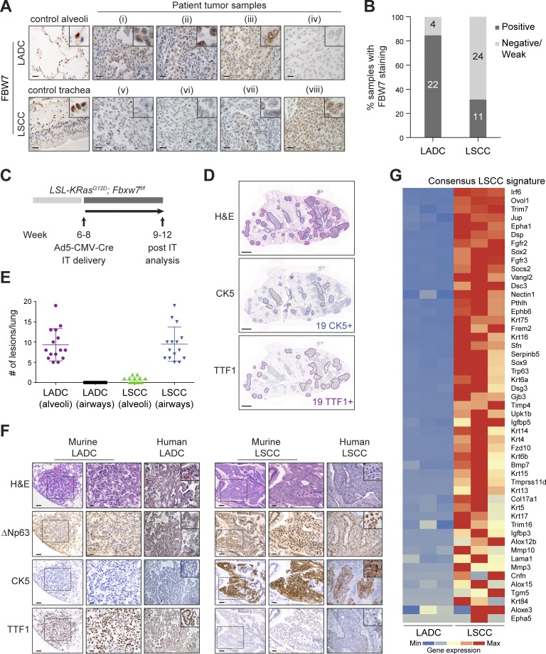
Figure 1.
Biallelic inactivation of Fbxw7 and KRasG12D activation in the adult mouse lung leads to LSCC and LADC formation. (A) Representative human lung LADC (i–iv) and LSCC (v–viii) tumors and control lung sections stained with FBW7 antibodies. Bars, 20 µm. (B) Quantification of FBW7 protein staining in human LADC and LSCC tumors as in A. n = 26 LADC, 35 LSCC. (C) Biallelic inactivation of Fbxw7 and KRasG12D activation by intratracheal (IT) delivery of Ad5-CMV-Cre virus in the adult mouse lung as a model of NSCLC. (D) KF model develops LSCC (CK5+) and LADC (TTF1+) tumors. Sections representative of six animals. (E) Quantification and localization of mouse lung LADC and LSCC tumors in the KF model. n = 15 lungs. Plots indicate mean ± SD. (F) Human and mouse NSCLC samples were stained with biomarkers used clinically to distinguish LADC (TTF1) from LSCC (CK5 and ΔNp63) tumors. Bars, 100 µm (columns 1 and 4); 20 µm (columns 2 and 5). Sections representative of six animals. (G) Heat map of RNASeq data showing relative expression of LSCC genes in LADC and LSCC tumors from n = 3 mice of the KF genotype. Gene set shown is selected from gene sets up-regulated in Lkb1f/f; Ptenf/f LSCC (Xu et al., 2014) and in LSL-Sox2; Ptenf/f; Cdkn2abf/f LSCC and human LSCC (Ferone et al., 2016). Genes are ordered according to z-score. See also Fig. S1.

