Figure 2.
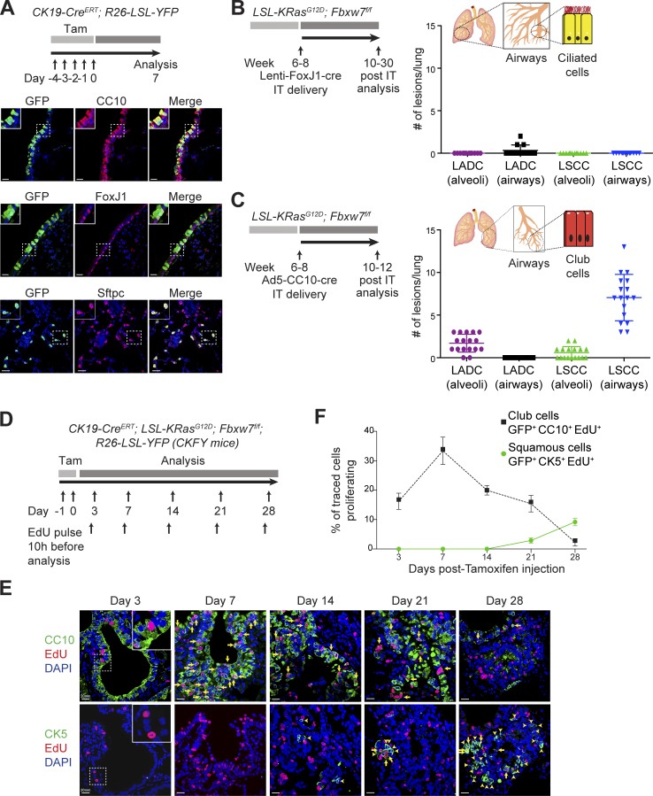
Lung LSCC originates from CC10+ luminal cells in the KF model. (A) Immunofluorescent staining for CC10, FoxJ1, Sftpc, and GFP in CK19-CreERT; R26-LSL-YFP mouse lung sections at 1 wk after induction. Images representative of three animals. Bars, 20 µm. Tam, tamoxifen. (B) Ciliated FoxJ1+ cells were targeted by intratracheal (IT) delivery of Lenti-FoxJ1-Cre virus to KF animals. The graph shows the quantification and localization of lung tumors produced. n = 12 lungs. Plots indicate mean ± SD. (C) Club CC10+ cells were targeted by intratracheal delivery of Ad5-CC10-Cre virus to KF animals. The graph shows the quantification and localization of lung tumors produced. n = 17 lungs. Plots indicate mean ± SD. (D) Scheme for analysis of cell proliferation in CK19-CreERT; KRasG12D; Fbxw7f/f; R26-LSL-YFP (CKFY) mice. (E) Lung serial sections from CKFY mice were immunostained for CC10, CK5, and EdU at 3, 7, 14, 21, and 28 d after tamoxifen injection. Arrows indicate proliferating cells. Arrowheads indicate double CK19+CK5+ cells. Images representative of five animals. Bars, 20 µm. (F) Quantification of proliferating CC10+ and CK5+ cells at the indicated time points. Graph indicates mean ± SD of five animals. See also Figs. S1, S2, and S3.

