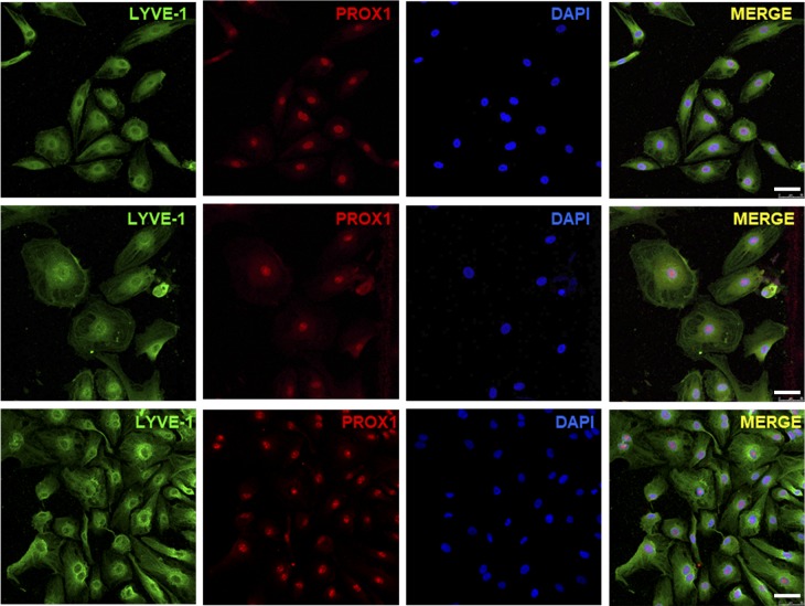
Figure 3.
Immunofluorescence staining of LECs isolated from patients with GLA. Representative immunofluorescence images of LECs isolated from patients with GLA (GLA0054 and GLA0061), compared with commercial human dermal LECs, stained with specific markers (LYVE-1 and PROX1) and analyzed by confocal microscopy. Bars, 50 µm.

