Figure 1.
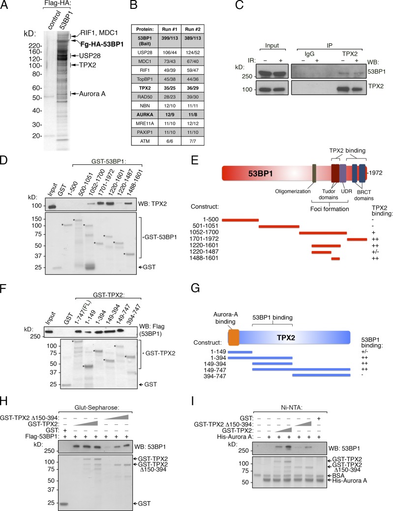
Identification of TPX2/Aurora A as 53BP1 effectors. (A) Flag-HA-53BP1 and associated proteins were isolated from HeLa-S nuclear extract using sequential Flag and HA immunoaffinity purification. The final eluted material was analyzed by SDS-PAGE and silver staining. (B) The 53BP1 complex from A was analyzed by MS/MS twice, and the total/unique peptide numbers for each protein are shown for each run. (C) Nuclear extracts from HeLa-S cells were immunoprecipitated with control IgG or TPX2 antibody and Western blotted as shown. (D) GST, or the indicated GST-53BP1 fragments, were immobilized on glutathione-Sepharose, and binding with full-length recombinant His-TPX2 was tested. Bound material was analyzed by Western blot or Coomassie Blue staining. (E) Schematic of 53BP1 and summary of TPX2 binding data. (F) GST, GST-TPX2, or the indicated GST-TPX2 fragments were immobilized on glutathione-Sepharose and assessed for binding to full-length Flag-53BP1, as in D. (G) Schematic of TPX2 and summary of 53BP1 binding data. (H) GST, and increasing equimolar amounts of GST-TPX2 or GST- TPX2-Δ150-394, were tested for 53BP1 binding as done in F. (I) Recombinant His-Aurora A was immobilized on Ni-NTA and incubated with GST, GST-TPX2, or GST- TPX2-Δ150-394. Bound material was washed, subsequently incubated with Flag-53BP1, washed again, and analyzed for binding by Western blot or Coomassie Blue staining.

