Figure 2.
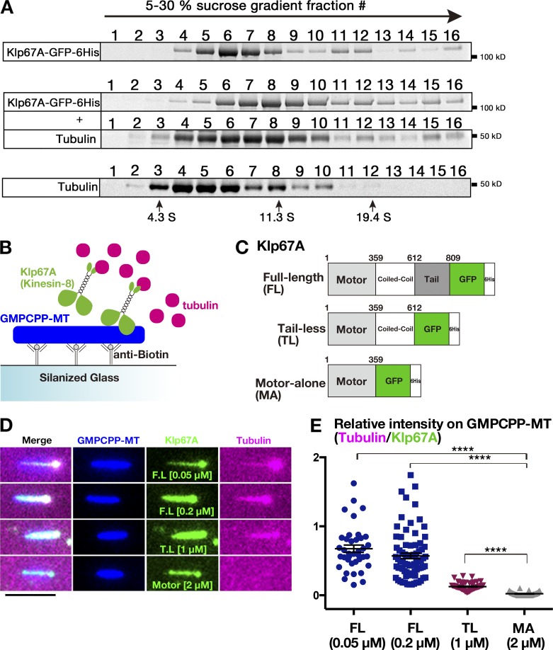
Tubulin-binding activity of kinesin-8Klp67A. (A) Co-fractionation of kinesin-8Klp67A-GFP and tubulin after sucrose gradient centrifugation. Each fraction was subjected to SDS-PAGE, followed by staining with Sypro Ruby. (B–D) Tubulin recruitment by kinesin-8Klp67A. Tubulin (magenta; 10 µM) and kinesin-8Klp67A (green; full-length, tail-less [1–612 aa], motor-alone [1–359 aa]), which bound to GMPCPP-stabilized MTs (blue), were mixed, and tubulin localization along MTs was investigated. Bar, 5 µm. (E) Quantification of tubulin intensity on the MT seed. Each dot represents a value obtained from a single MT and error bars represent SEM. FL versus MA: P = 3.8 ×10⁻¹⁴, and TL versus MA: P = 3.8 ×10⁻¹⁰ by Games-Howell test. n = 38 (50 nM), 77 (200 nM; full length), 33 (motor alone), and 58 (tail-less).

