Figure 2.
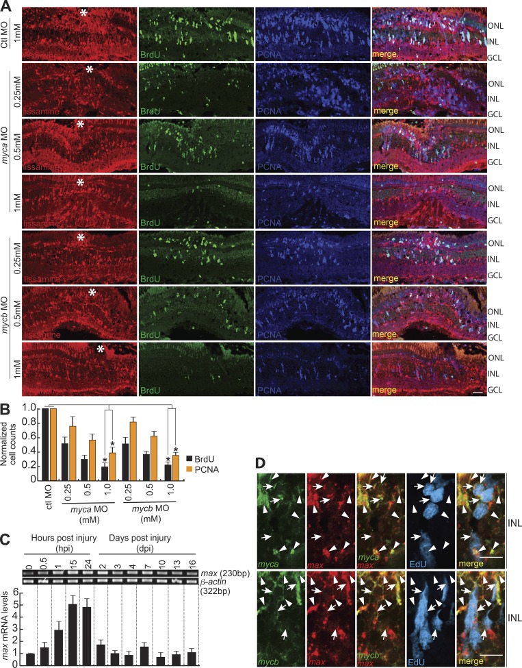
Myc is necessary for MG dedifferentiation in the injured retina. (A) IF microscopy images of control (Ctl; 1 mM concentration) or myca/mycb-targeting lissamine-labeled MOs (0.25, 0.5, and 1 mM concentration each), electroporated into the retina of zebrafish at the time of retinal injury shows a concentration-dependent decrease in the number of MGPCs. Fish were given an intraperitoneal injection of BrdU, 3 h before euthanasia on 4 dpi. The white asterisks mark the injury sites. (B) Quantification of the number of BrdU+ and PCNA+ cells at the injury site. The data are compared with control MO. *, P < 0.001; n = 4 biological replicates. (C) RT-PCR (top) and qPCR (bottom) were used to assay injury-dependent max gene expression; n = 6 biological replicates. (D) ISH and IF microscopy show that max gene expression colabel with myca and mycb mRNA in EdU+ MGPCs and other surrounding cells at 4 dpi. White arrowheads indicate myca or mycb colabeled with max, and white arrows mark myca/mycb/max in EdU+ MGPCs. Bars, 10 µm (A and D). Error bars are SD. ONL, outer nuclear layer; INL, inner nuclear layer.

