Figure 3.
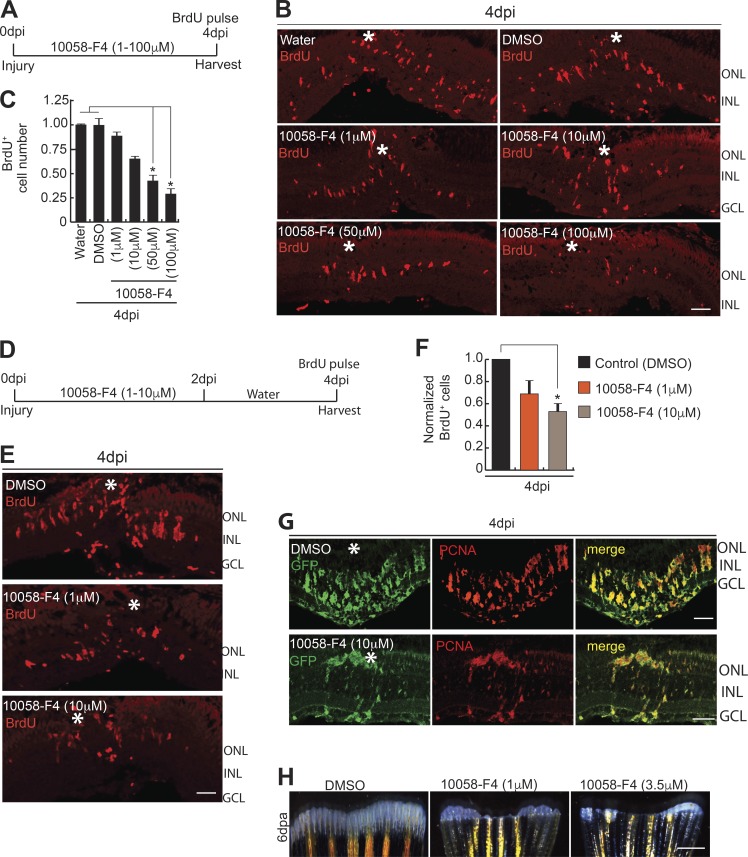
Blockade of Myc–Max interaction abolishes MGPC proliferation in retina and fin regeneration. (A–C) Blockade of the Myc–Max interaction using the drug 10058-F4 treatment, as shown in timeline of experiment (A), reveals significant reduction in BrdU+ MGPCs at 4 dpi, seen by IF microscopy (B), which is quantified and normalized to Water/DMSO control (C). *, P < 0.0001; n = 5 biological replicates. (D–F) Early treatment with 10058-F4, as shown in timeline of experiment (D), also reveals a significant reduction in BrdU+ MGPCs at 4 dpi revealed by IF microscopy (E), which is quantified and normalized to DMSO control (F). *, P < 0.01; n = 5 biological replicates. (G) IF microscopy analysis of GFP and PCNA after 10058-F4 treatment shows a reduction in the number of MGPCs in 1016 tuba1a:gfp transgenic fish retina at 4 dpi. (H) Regenerating fin-blastema shows a decline in cell mass in 10058-F4–treated post-amputated fin (1 µM and 3.5 µM) at 6 d after amputation, compared with DMSO control. Error bars are SD. Bars: 10 µm (B, E, and G) and 500 µm (H). White asterisks mark the injury sites (B, E, and G). ONL, outer nuclear layer; INL, inner nuclear layer.

