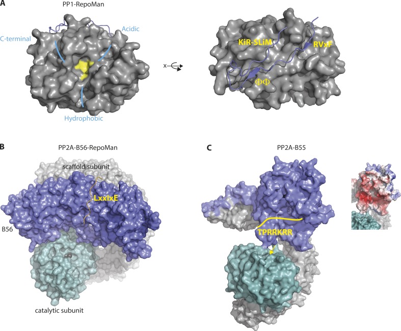
Figure 2.
Structural aspects of mitotic phosphatases and binding to SLiMs. (A) Structure of PP1 in complex with RepoMan. The active site (yellow), the three possible substrate-binding grooves around the active site (light blue), and the binding of RepoMan motifs to different pockets on PP1 are indicated. (B) Model of PP2A-B56 bound to the LxxIxE motif of RepoMan with catalytic subunit (turquoise), scaffold (gray), and B56 (blue). (C) Structure of PP2A-B55 with the hypothetical binding of a basic region to the acidic region on the B55 subunit. The electrostatic potential of the B55 surface is shown on the right with basic residues in red. The yellow arrow indicates the active site.

