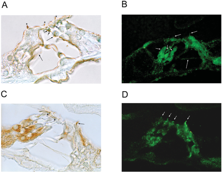
Figure 7. Immunolocalization of EMILIN1 and PDE6C in rat organ of Corti.
(A) EMILIN1 was immunolocalized with DAB to nerve fibres (efferents) crossing the tunnel of Corti ending at the base of outer hair cells (long arrows) in rat organ of Corti. Small nerve fibres at subcuticular plate sites of outer hair cells were immunoreactive (short arrows) and stereociliary arrays of both inner and outer hair cells exhibited immunoreactivity for EMILIN1 (asterisks). (B) With fluorescence detection, EMILIN1 immunoreactivity was again found in olivocochlear axonal efferent fibres crossing the tunnel (long arrow), making contact with the base of the outer hair cells and Deiters’ cells (short arrows). Immunoreactivity was clearly associated with stereocilia of both inner and outer hair cells (mid-length arrows). (C) PDE6C immunoreactivity was associated with Deiters’ cells; however, this was not due to overlapping efferent/afferent contacts, since neither the tunnel crossing efferents nor the type II afferents were immunoreactive. PDE6C immunoreactivity was found at subcuticular sites on outer hair cells (short arrows) and was strongly present on stereocilia of the inner hair cell (mid-length arrow). (D) PDE6C immunofluorescence was associated with stereocilia of both inner and outer hair cells (arrows).

