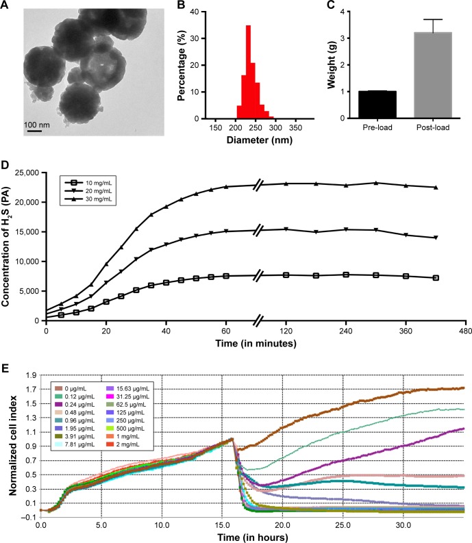
Figure 1.
Characterization of DATS-MIONs.
Notes: (A) TEM imaging of a single nanoparticle. The scale bar is shown in the lower left corner. (B) Size distribution of DATS-MIONs. (C) Drug loading experiment. One gram of MIONs was used, and the weight of the loaded nanoparticles is shown. (D) Time course of H2S release by DATS-MIONs at different nanoparticle concentrations measured on an H2S-selective microelectrode. (E) Proliferation of HUVECs incubated with different concentrations of DATS-MIONs as monitored on a real-time cell analyzer.
Abbreviations: DATS, diallyl trisulfide; HUVECs, human umbilical vein endothelial cells; MIONs, mesoporous iron oxide nanoparticles; TEM, transmission electron microscopy.

