Figure 5.
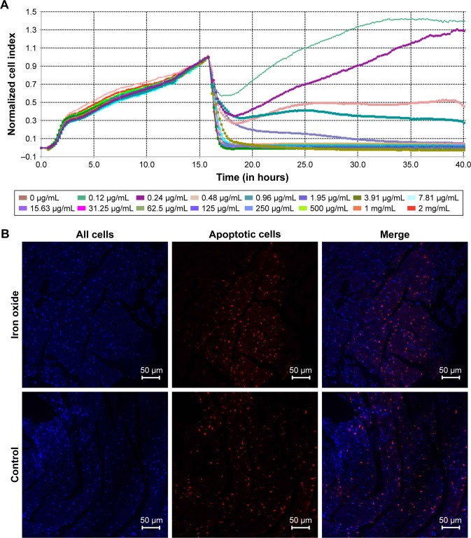
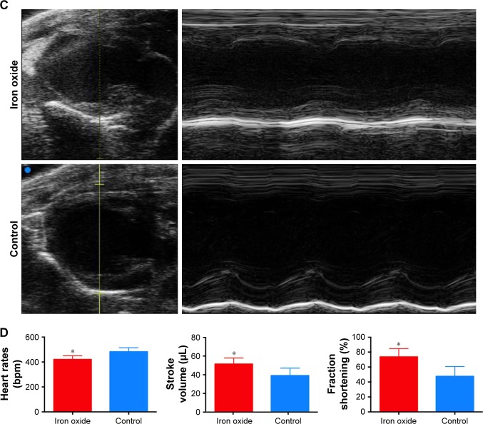
Evaluation of the effect of DATS-MIONs on myocardial protection.
Notes: (A) Proliferation of embryonic cardiomyocyte H9C2 cells incubated with different concentrations of DATS-MIONs as monitored on a real-time cell analyzer. (B) TUNEL assay of myocardial tissues harvested from treated (iron oxide, top) and untreated (control, bottom) mice with IR-induced heart injury. Apoptotic cells were stained positive in the assay and highlighted by the red fluorescence that they emitted. (C) Representative echocardiograms of a mouse with MI in which DATS-MIONs were injected and a mouse with MI as a control that did not receive any treatment. (D) Column charts comparing the heart rate, ejection fraction, and fraction shortening of the three abovementioned murine models. Bars represent standard error of mean. *P<0.05.
Abbreviations: DATS, diallyl trisulfide; IR, ischemia–reperfusion; MI, myocardial infarction; MIONs, mesoporous iron oxide nanoparticles.


