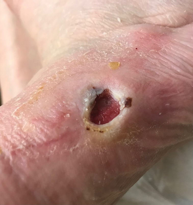
Fig. 5.

Neuropathic ulcer. Note the peri-wound callus, well-defined edges (“punched-out” appearance), red wound bed, no fibrinous exudate, and the location on the lateral aspect of the left foot of the same patient as in Fig. 4 , an abnormal pressure point due to Charcot deformity.
