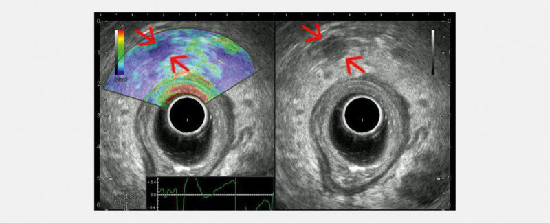
Fig. 3.

Radial endorectal ultrasound (ERUS) elastography showing a hard (low strain) perirectal lymph node (red arrows) in a patient with concomitant rectal adenocarcinoma. A balloon surrounding the transducer is inflated with water to improve acoustic coupling with the rectal wall (courtesy of Adrian Săftoiu, Elena Tatiana Ivan).
