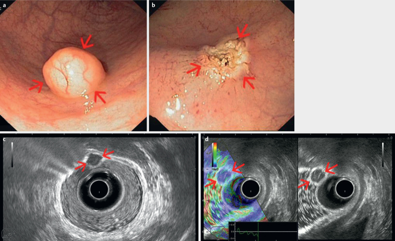
Fig. 7.

ab Early rectal neuroendocrine tumor visualized endoscopically as a small mass with normal appearing mucosa, completely resected by endoscopic mucosal resection. cd Radial endorectal ultrasound (ERUS) delineates the small tumor (red arrows) as a hypoechoic mass, hard by elastography, limited to the mucosa, with clear demarcation from the submucosa and muscularis propria (T1). Water has been instilled in the balloon covering the ultrasound transducer, as well as in the rectum for better acoustic coupling between the transducer and rectal structures (courtesy of Adrian Săftoiu, Elena Tatiana Ivan).
