
Fig. 8.
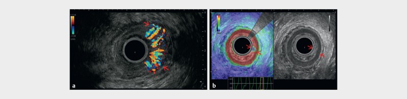
a Color flow imaging showing the Doppler appearance of a hypervascular inflammed anal region. b Endoanal ultrasound (EAUS) with a 360-degree radial transducer showing the muscular layers of the anal canal (red arrows), as well as the soft appearance of an inflammed anal sphincter region (courtesy of Christoph F. Dietrich).