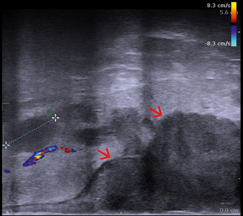
Fig. 10.

Linear endoanal ultrasound (EAUS) showing the tumor (red arrows) extension from the anodermal junction, as well as thickness, with an oval, hypoechoic, well demarcated lymph node of 15 mm in the perirectal fat shown between markers (uT3N1) (courtesy of Christian Nolsøe, Torben Lorentzen).
