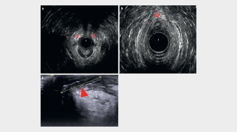
Fig. 15.

a Anterior half of internal sphincter (IS) defect (red arrows) visualized by radial endoanal ultrasound (EAUS). b Radial EAUS of an anteriorly located fistula (between calipers), with an endoanal image (360°) showing 12 o’clock inter-sphincteric portion of the fistula (red arrow). c Perineal ultrasound (PNUS) view with a high frequency linear transducer depicts well the other portion of the fistula in between calipers (arrowhead), which is out of field of view for endoanal image (courtesy of Ismail Mihmanli).
