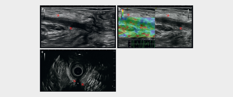
Fig. 20.

a Trans-sphincteric fistula (red arrows) visualized with perineal ultrasound (PNUS) in the extrasphincteric course. b PNUS elastography showing the soft (compressible) fistula tract (red arrows). c Endoanal ultrasound (EAUS) appearance of the rest of the fistula tract (red arrow) (courtesy of Christoph F. Dietrich).
