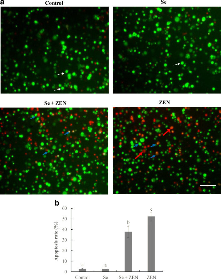
Fig. 3.
Observation of morphological apoptosis in chicken splenic lymphocyte treated with ZEN and Se. a Fluorescence images determined by staining with acridine orange and ethidium bromide (AO/EB). The cells were treated with control (untreated), Se, Se + ZEN, and ZEN. White indicate arrows live cells; blue arrows indicate apoptotic cells; red arrows indicate necrotic cells

