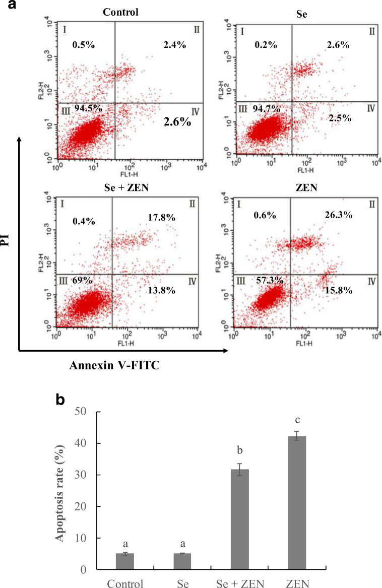
Fig. 4.
Changes of apoptosis in chicken splenic lymphocyte treated with ZEN and Se. Apoptosis was evaluated by flow cytometry using annexin V/PI staining. a The cells were treated with control (untreated), Se, Se + ZEN and ZEN. Quadrants: I, necrotic cells; II, late apoptosis cells; III, living cells; and IV, early apoptosis cells. b Columns, mean of three experiments, and the data represent the mean ± SEM; different lowercase letters indicate that there are statistically significance (P < 0.05) among different groups

