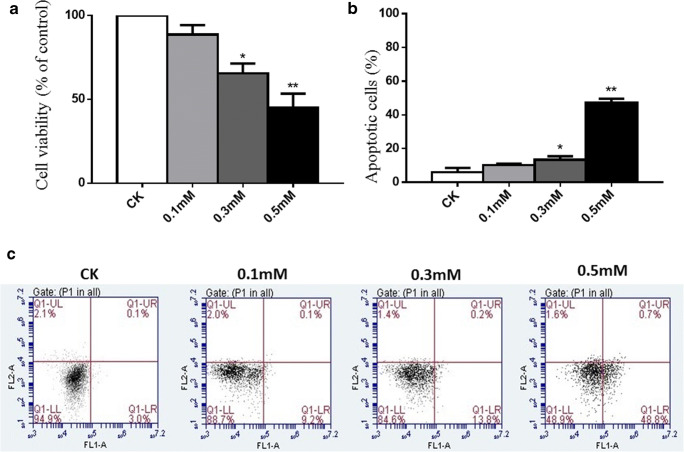
Fig. 1.
Cell viability and cell apoptosis in chondrocytes treated with different concentrations of H2O2. a Normal chondrocytes (CK) were treated with different concentrations of H2O2 (0.1 mM, 0.3 mM, and 0.5 mM) for 30 min. Cell viability was detected by CCK-8. b Results of cell apoptosis in different groups. c Normal chondrocytes (CK) were treated with different concentrations of H2O2 (0.1 mM, 0.3 mM, and 0.5 mM) for 30 min. Normal chondrocytes cultured in DMEM for 30 min were used as a negative control. FITC annexin V/PI staining and flow cytometry assays were used to detect cell apoptosis. Results are presented as means ± standard deviation of three independent experiments. Untreated chondrocytes were used as control and considered 100% viable. Asterisk indicates P < 0.05, double asterisks indicate P < 0.01 versus normal control

