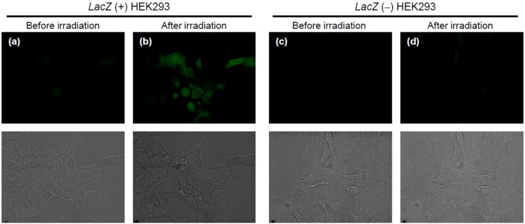
Figure 9.
Fluorescence imaging of NO release from NO-Rosa-Gal in lacZ-HEK293 cells using DAF-FM DA. Cultured HEK293 cells were treated with DAF-FM DA (10 μM) and NO-Rosa-Gal (10 μM). The dishes were then photoirradiated with blue light (530−590 nm, 84 mW/cm2 for 15 min). The dishes were observed with a confocal fluorescence microscope. Upper figures are green fluorescence, and bottom figures are bright filed. (a) before photoirradiation to lacZ (+) cells, (b) after photoirradiation to lacZ (+) cells, (c) before photoirradiation to lacZ (−) cells, (d) after photoirradiation to lacZ (−) cells.

