Figure 4.
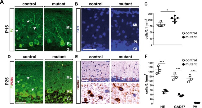
NeuroD2 is required for survival and terminal differentiation of MLIs. (A) Absence of PV+ MLI in the cerebellum of NeuroD1−/− mutants. Fluorescent immunostaining of sagittal sections (lobule 5) at P15. Arrowheads point to PV+ MLIs in control mice. Scale bar, 50 µm. (B) Nuclear DAPI staining of sagittal sections (lobule 5) at P15. Scale bar, 50 µm. (C) Increased density of DAPI+ nuclei in the ML of NeuroD−/− mutants. n = 4–5 per genotype. (D) Fluorescent immunostaining of sagittal sections (lobule 5) at P25. Arrowheads point to PV+ MLIs in control mice. Scale bar, 50 µm. (E) H&E staining and immunostaining for GAD67 on sagittal sections (P25, lobule 5). (F) Reduced density of GAD67+ interneurons in the ML of NeuroD2−/− mutants. Quantification of H&E and GAD+ stainings in lobule 5, n = 3 per genotype.

