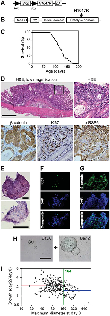
Figure 3. Fc1 Apcfl/+ Pik3caH1047R mice develop invasive colon adenocarcinomas and can be utilized for translational investigations.
Fc1 Apcfl/+ Pik3caH1047R mice possess a lox-stop-lox sequence prior to the human PIK3CA H1047R hotspot mutation (A and B). These mice were allowed to age until moribund. These mice became moribund at an average age of 121 days (C). At necropsy tumors were identified within the small intestine and colon. Upon histologic sectioning, these tumors were found to be well to moderately differentiated adenocarcinomas (D, upper panels). These cancers possessed nuclear CTNNB1 (β-catenin), increased Ki67 and phosphorylation of RPS6 (D, lower panels). Spheroid cultures were able to be generated from these cancers and demonstrated a similar histology (E) and proliferating cells as measured by Ki67 (F). Nuclear CTNNB1 was also observed in the spheroids (G). The diameter of these spheres can be followed over time as a marker of proliferation (H). It was observed that larger spheroids at baseline would typically grow at a slower rate. To standardize experiments between cohorts, a standard change-point analysis was utilized (I). A change point at 164 pixels (373 μm) was identified and only those spheroids below that cut-off were utilized in further investigations. Size bars: D = 500 μm, E = 100 μm, G = 1 mm. The square panels in D are 4x enlargements of the outlined area.

