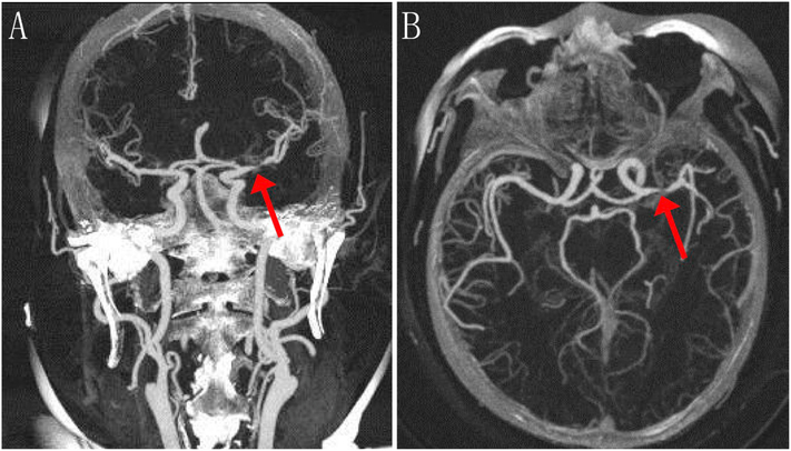
Fig. 2.
Representative CTA images of ischemic stroke with stenosis. Coronal (a) and axial (b) CTA images were taken 4 days after left MCA infarction in a 55-year-old woman with right hemiparesis and aphasia. A high-grade stenosis at the proximal left MCA (M1 segment) is denoted with red arrows. (For interpretation of the references to colour in this figure legend, the reader is referred to the web version of this article.)

