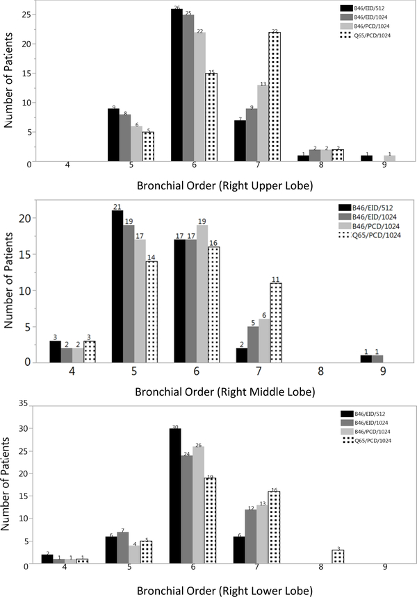
Figure 1.
Bar plots of the frequency of higher (more peripheral) bronchial order detection for both readers in right upper (a), middle (b), and lower (c) lobes by CT acquisition and image reconstruction method. Higher order bronchi were detected in all right lung lobes for the PCD-CT images with 1024 ×1024 matrices regardless of the kernel used (light grey (B46), patterned (Q65) bars) compared to clinical reference (black bar) (a-c). The addition of the Q65 sharp kernel (patterned bars) increased the detection of 7th and 8th order bronchi compared to all other CT acquisition and image reconstruction methods.

