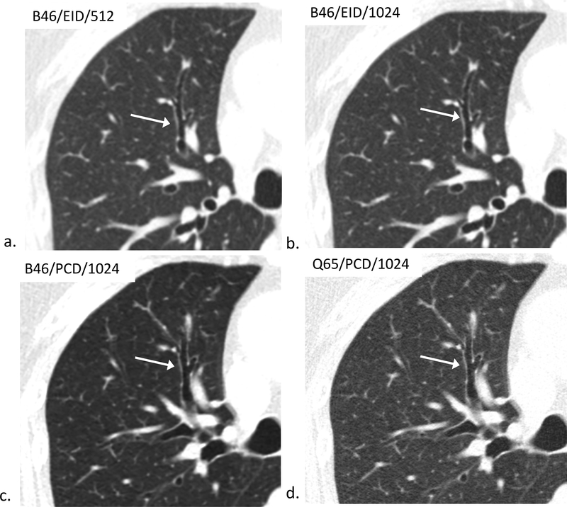
Figure 4.
80-year-old female evaluated for hemoptysis. B46/EID/512 (a), B46/EID/1024 (b), B46/PCD/1024 (c), Q65/PCD/1024 (d) images demonstrating improved visualization of the same normal 4th order bronchial wall in the Q65/PCD/1024 (d) image compared to clinical reference (a). The readers mean evaluation scores for 4th order bronchial wall evaluations were 0.5 (b), 0.5 (c), and 1.5 (d), respectively; W/L = 1200/−600 HU.

