
Figure 2.
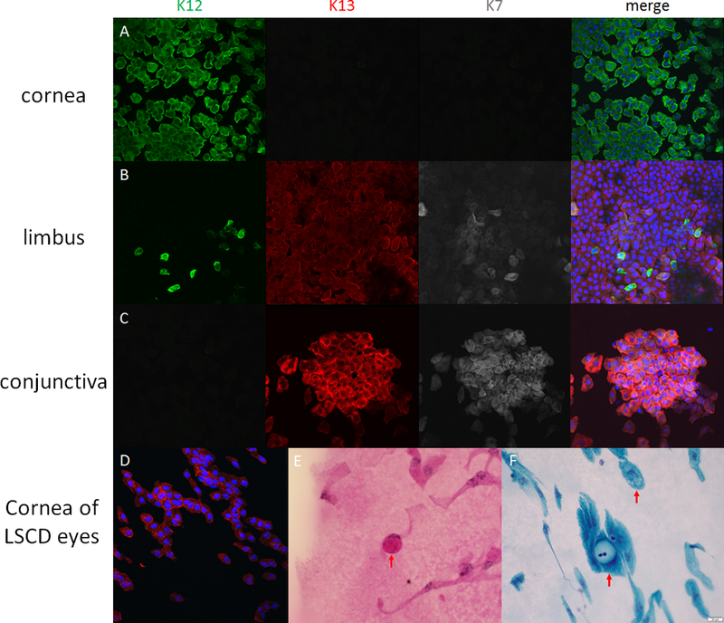
Immunofluorescein and pathological staining of corneal impression cytology specimens. Figure 2A, 2B and 2C are specimens taken from central cornea, limbus and conjunctiva of a normal cadeveric eye, respectively. The expression of cytokeratin (CK) 12 is only found in corneal epithelial cells, and cannot be found in conjunctival epithelial cells. CK13 and CK7 are only expressed in conjunctival cells. At the limbal area, both CK12-positive cells and CK13-CK7-positive cells are visible. Figure 2 D-F are the samples taken from the eyes with limbal stem cell deficiency. CK13 positive cells are visible on the cornea (2D). The goblet cells can be found through periodic acid-Schiff staining (2E) or Papanicolaou staining (2F) in the samples taken in limbal stem cell deficiency eyes after immunostaining. The goblet cells have 2–3 times larger cell size compared with epithelial cells, and mucus blobs are visible within the cells. The mucus blobs are usually visualized as pink by PAS staining.