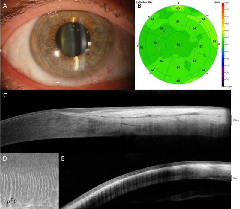
Figure 4.

AS-OCT assessment of normal eyes (A-E) and eyes with limbal stem cell deficiency (LSCD, F-J). In eyes with sectoral LSCD, the corneal epithelial thickness in the affected area is decreased (G) compared to normal eye (B) and unaffected area (G); this reduction is consistent with the slit-lamp finding (F). Cross-sections perpendicular to the limbus show a clear transition between the hyporeflective corneal epithelium and the hyperreflective conjunctival epithelium with limbal epithelial thickening in normal eyes (C) but not in eyes with LSCD (H). The limbal epithelium in the affected area becomes thinner (H). The palisades of Vogt (p in figure) and limbal crypts (c in figure) are clearly visualized in normal eyes on en face mode (D) and parallel section (E), whereas they are absent in LSCD eyes (I and J).
