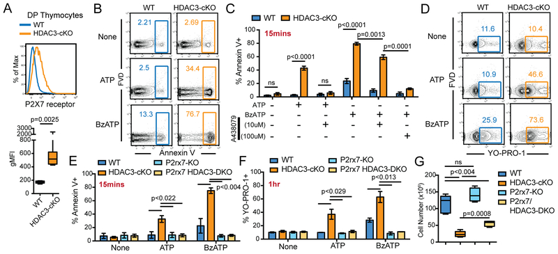
Figure 2. HDAC3-deficient DP thymocytes are susceptible to cell death mediated by the P2X7 receptor.
(A) P2X7 receptor expression on DP thymocytes from 5 WT and 7 HDAC3-cKO mice from 3 independent experiments. (B-C) Frequency of Annexin V+ DP thymocytes stimulated for 15 minutes ex vivo with 1mM of ATP or 100μM of BzATP from WT and HDAC3-cKO mice, with or without a 1 hour pre-treatment with the P2X7 receptor antagonist A438079. Plots show mean ± SEM of 3–4 mice per group from 3 independent experiments. (D) Frequency of YO-PRO-1+ DP thymocytes, from WT and HDAC3-cKO mice, stimulated for 1 hour ex vivo with 1mM ATP or 100μM BzATP. Data is representative of 3–4 mice from 3 independent experiments. (E-F) Frequency of DP thymocytes that are Annexin V+ (E) or YO-PRO-1+ (F) after ex vivo stimulation with 1mM of ATP or 100μM of BzATP for 15 minutes or 1 hour, respectively. DP thymocytes are from WT, HDAC3-cKO, P2rx7-KO, or P2rx7/HDAC3-DKO mice. Plot shows mean ± SEM of 3–4 mice from 3 independent experiments. (G) Number of DP thymocytes from WT, HDAC3-cKO, P2rx7-KO or P2rx7/HDAC3-DKO mice. Box plots depict 4–5 mice from 4 independent experiments.

