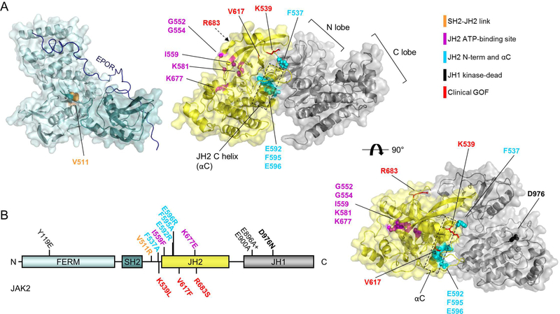
Figure 1: JAK2 domain structure.
A: Structures of JAK2 FERM-SH2 (left, PDB: 4Z32) with model of EPOR JAK2-binding peptide shown in dark blue (modelled based on Interferon λ 1 receptor (IFNLR1) peptide bound to JAK1 FERM-SH2, PDB: 5L04), and JAK2 JH2-JH1 inhibitory interaction 12. Right: JAK2 JH2-JH1 top view. B: Domain structure of JAK2. See also Table 1.

