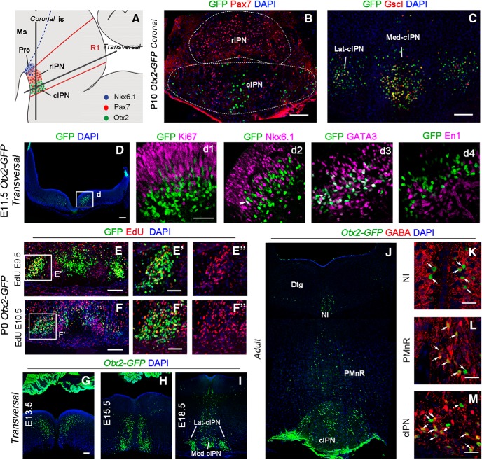
Figure 5.
Otx2 expression pattern in the IPN. A, Schematic illustration of a sagittal section of IPN subnuclei in the ventral r1 in embryonic brain. B, Coronal section of P10 Otx2-GFP mouse brain at the level of the IPN. The rostral IPN is characterized by high density of Pax7 neurons. Otx2 neurons (GFP) in the cIPN form two subnuclei, the medial and the lateral one, with some Pax7 neurons in between. C, Coronal section of the IPN showing colocalization between Otx2 and Gscl in the cIPN. D, Transversal section of E11.5 Otx2-GFP embryo at the level of the r1 showing Otx2 neurons in the basal plate. Dd1–Dd4, High magnification of the box area indicated in D showing potential Otx2 neurons (GFP+) in r1 that also contain Ki67 (Dd1), Nkx6.1 (Dd2), GATA3 (Dd3), and En1 (Dd4) at embryonic stage E11.5. E, F, High magnification of the IPN area on P0 Otx2-GFP embryo showing colocalization between Otx2 (GFP+) and Edu injected at E9.5 (E) and E10.5 (F). E′–F′′, High magnification of the boxed area. G–M, Immunolabeling in transversal section of the r1 in Otx2-GFP embryos showing the distribution of Otx2 neurons (GFP+) in the basal plate at different embryonic (G–I) and adult stages (J–M). K–M, Colocalization between Otx2 and GABA in the nucleus incertus, PMnR, and cIPN. Ms, Mesencephalon; is, isthmus; Pro, prodromal division of the IPN; Dtgm dorsal tegmental nucleus; NI, nucleus incertus. Scale bars, B–D and G–I, 100 μm; Dd1–Dd4 (shown in Dd1) and E′–F′′ (shown in E′ and F′), 50 μm; K–M, 25 μm.

