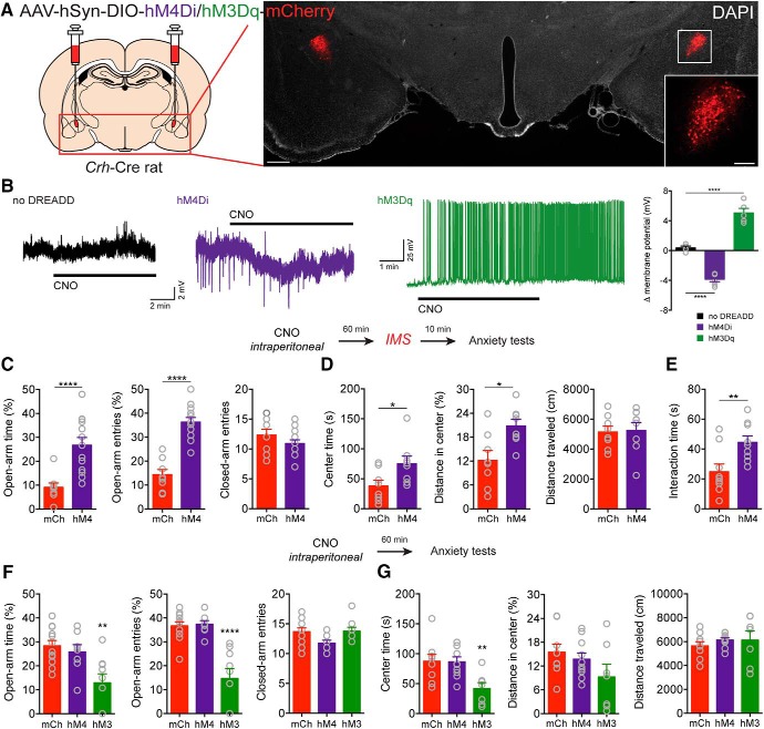
Figure 1.
CRFCeA neurons contribute to stress-induced anxiety. A, Injection schematic and image of AAV-DREADD-mCherry expression in CRFCeA neurons. Scale bar, 500 μm. Boxed region is enlarged in inset. Scale bar, 200 μm. B, Representative traces showing that bath application of CNO (2 μm) had no effect in a neuron not expressing a DREADD (left), induced hyperpolarization of a neuron expressing DIO-hM4Di (middle), and promoted depolarization and spontaneous firing in a neuron expressing DIO-hM3Dq (right). Far right, Quantification of changes in resting membrane potential in CeA neurons during CNO application. C, CNO (2 mg/kg, i.p.) increased the percentage of time spent in the open arms of the EPM and the percentage of open arm entries without changing the number of closed arm entries. D, Activation of hM4Di with CNO increased the time spent in the center and the distance traveled in the center of the OF but did not alter the total distance traveled. E, CNO also increased social interaction time in rats that expressed hM4Di compared with control rats that expressed mCherry in CRFCeA neurons. F, In rats that expressed hM3Dq in CRFCeA neurons, CNO (2 mg/kg, i.p.) reduced the percentage of time spent on the open arms of the EPM and reduced the percentage of open arm entries without affecting the number of closed arm entries. G, hM3Dq reduced the time spent in the center of the OF and marginally reduced the distance traveled in the center of the OF without affecting the total distance traveled. In contrast, activation of hM4Di had no effect on anxiety-like behavior when compared with mCh control animals. Data are mean ± SEM. *p < 0.05, **p < 0.01, ****p < 0.0001. mCh (mCherry), hM4 (hM4Di), hM3 (hM3Dq).

