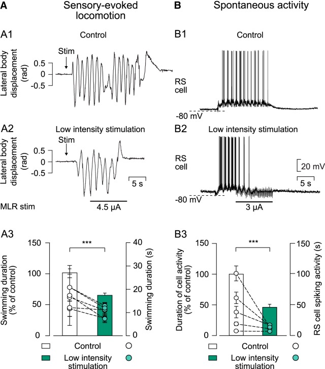
Figure 4.
Effect of a low-intensity MLR stimulation on ongoing sensory-evoked or spontaneous swimming. A1, Kinematic analysis of the lateral body displacement (rad) during sensory-evoked swimming that was elicited by pinching the dorsal fin with forceps (Stim). A2, Representation of sensory-evoked swimming when a low-intensity stimulation was delivered to the MLR 5 s after the onset of swimming. A3, Histogram illustrating pooled data of average swimming duration (n = 30 trials in 6 animals) in control conditions (white bar) and when MLR was stimulated electrically of low intensity during sensory-evoked swimming (green bar). B1, Intracellular recording of a maintain cell that fires action potentials throughout the locomotor bout (monitored visually) used to analyze spontaneous locomotor activity. B2, Representation of cellular activity when MLR stimulation of low intensity was delivered during spontaneous swimming. B3, Histogram illustrating pooled data of duration of cellular activity in 5 animals (n = 25 events) in control conditions (white bar) and when the MLR was stimulated 5 s after swimming movements had started (green bar). In both histograms, bars represent mean ± SEM of the duration of swimming episodes or cellular activity normalized to average value of control (left y-axis). Dots represent average duration of swimming episodes or cellular discharge for each animal (mean ± SEM; right y-axis). In all experiments, MLR stimulation intensities that would not induce locomotor activity in the resting preparation were used. ***p < 0.001.

