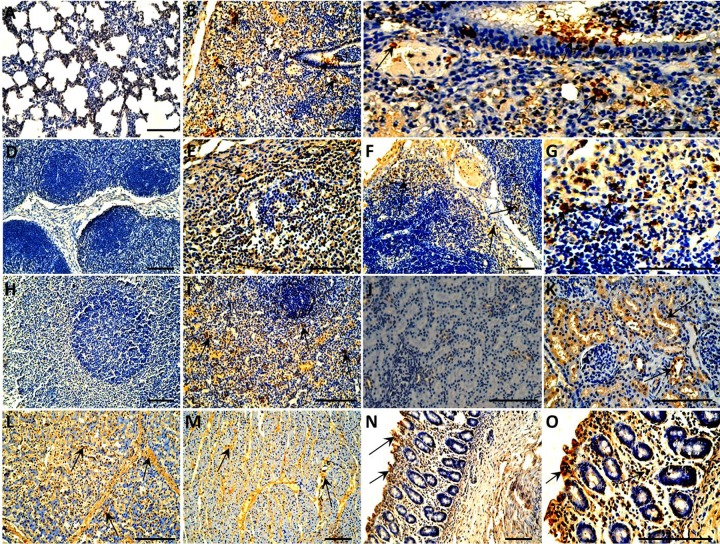
FIG 7.
Immunohistochemical staining of organs and tissues of the 4-week-old piglets inoculated with PCV3. PCV3 antigen-positive cells are brown. (A) No staining was observed in the lung section from a sham-inoculated piglet. (B) Lung tissues from a PCV3-inoculated piglet showed many cells positive for PCV3 antigen (arrows). (C) Partial enlargement of panel B. (D) No staining was observed in the lymphoid tissue section from a sham-inoculated piglet. Tracheobronchial lymph node (E) and mesenteric lymph node (F) from the PCV3-inoculated piglets show many cells positive for PCV3 antigen (arrows). (G) Partial enlargement of panel F. (H) No staining was observed in the spleen section from a sham-inoculated piglet. (I) Spleen from a PCV3-inoculated piglet showed many cells positive for PCV3 antigen (arrows). (J) No staining was observed in the kidney section from a sham-inoculated piglet. (K) Kidney from a PCV3-inoculated piglet showed many cells positive for PCV3 antigen (arrows). Many cells positive for PCV3 antigen (arrows) were also observed in liver (L), heart (M), and small intestine (N) of a PCV3-inoculated piglet. (O) Partial enlargement of panel N. Bars, 80 μm.

